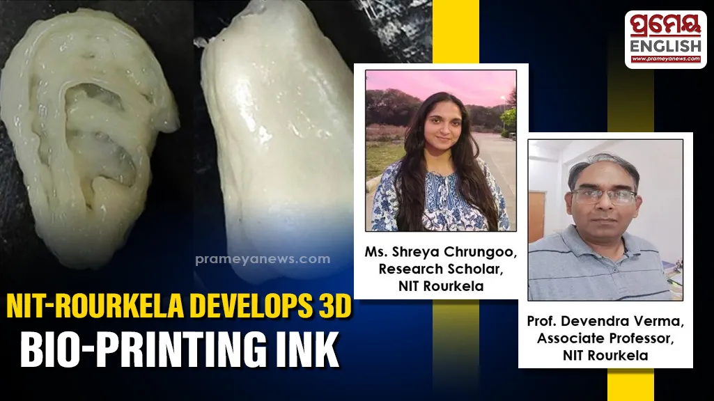
Rourkela, April 20: National Institute of Technology Rourkela (NIT Rourkela) research team has developed a novel bio-ink that can be used in 3D bioprinting and tissue engineering.
Bio-inks are materials used in 3D bioprinting to fabricate tissue-like structures. However, due to lack of bio-inks that combine mechanical strength, biological compatibility, and printability, broad usage of this technology remains limited.
To address this challenge, Prof. Devendra Verma, Associate Professor, along with his research scholar, Shreya Chrungoo, and Dr. Tanmay Bharadwaj, Dept. of Biotechnology and Medical Engineering, NIT Rourkela, have developed a high shape-fidelity protein–polysaccharide bio-ink that can be effectively used in bone and cartilage repairing.
The findings of this research have been published in the prestigious International Journal of Biological Macromolecules. The research team has also secured a patent titled, “AHigh Shape-Fidelity Protein-Polysaccharide Composite Bioink for 3DBioprinting” for the developed technology. (Patent number: 583759; Application number: 202431019470).
To achieve desired results, the research team combined Bovine Serum Albumin (BSA), Sodium Alginate, and Polyelectrolyte complexes of gelatin and chitosan (PEC-GC). The blend created a bioactive system that supported cell growth while maintaining structural fidelity during and afterthe printing process.

Speaking about the unique feature of developed bioink, Prof. Devendra Verma, said, “Our goal was to bridge the long-standing gap between printability and biological performance in bio-inks. By integrating protein–polysaccharide interactions with nanofibrous complexes, we have developed a system that not only prints with high precision but also actively supports cellular functions and tissue regeneration. This brings us a step closer to clinically relevant bioprinted constructs.”
On lab scale trials, the research team found the developed bio ink mimicking the extracellular matrix of bone tissue, providing sites for cell attachment, and promoting cell adhesion, proliferation, and overall biological response. Additionally, the printed scaffolds were found to have strong mechanical properties helping in retaining shape and functionality post printing.
Experiments have shown that scaffolds containing 2% PEC-GC achieved over 90% cell viability. Additionally, it demonstrated potential for bone tissue formation and collagen synthesis.
Speaking about real-world usability of the developed bio-ink, research scholar, Ms. Shreya Chrungoo, said, “The developed bio-ink offers a versatile platform for fabricating patient-specific scaffolds with precise geometry and biological functionality. Its ability to support high cell viability and tissue-like behavior makes it promising for applications in regenerative medicine.”
As the next step, the research team plans to undertake animal studies to further establish the safety and efficacy of the developed bio-ink, followed by clinical studies for validation.
The developed bio-ink holds significant potential for regenerative medicine. Its ability to enable the fabrication of patient-specific, tissue-like structures opens new avenues in personalised healthcare and therapeutic applications.